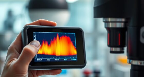

Fluorescence lifetime imaging (FLI) lets you photograph chemical reactions inside cells by measuring how long molecules emit fluorescence after excitation. This technique reveals changes in the molecular environment, such as pH or binding events, that signal reactions are occurring. By tracking these lifetime shifts, you can visualize dynamic processes in real time. Keep exploring, and you’ll discover how this approach provides detailed insights into cellular chemistry and helps you monitor reactions as they unfold.
Key Takeaways
- FLI captures changes in fluorescence decay times, revealing real-time chemical reactions and molecular interactions within living cells.
- Combining photoactivation with FLI allows selective tracking of molecules undergoing chemical transformations.
- Variations in fluorescence lifetime indicate environmental shifts, such as pH or binding events during cellular chemical reactions.
- Quantum dots provide stable, long-term fluorescence signals essential for monitoring dynamic chemical processes.
- FLI enables visualization and quantification of subtle biochemical reactions that are invisible to traditional intensity-based imaging.

Fluorescence lifetime imaging (FLI) is a powerful technique that measures the decay time of fluorescence emitted by molecules after excitation. This approach reveals details about the molecular environment and interactions within cells, enabling you to observe dynamic biological processes in real-time.
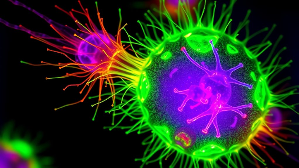
One exciting application of FLI involves quantum dot imaging, where you use semiconductor nanocrystals with unique fluorescence properties. Quantum dots have high brightness, stability, and size-tunable emission wavelengths, making them ideal for tracking multiple molecules simultaneously. When combined with fluorescence lifetime measurements, quantum dots can provide more precise information about their local surroundings, helping you distinguish between different cellular states or interactions that might be indistinguishable based on intensity alone.
Quantum dots enable multi-molecule tracking with enhanced environmental sensitivity via fluorescence lifetime measurements.
In addition to quantum dot imaging, photoactivation techniques markedly enhance your ability to study cellular processes with FLI. Photoactivation involves using specific wavelengths of light to activate or “turn on” fluorescent molecules in targeted regions within the cell. This allows you to selectively label and monitor particular molecules or structures over time.
When paired with fluorescence lifetime measurements, photoactivation enables you to track the movement and interaction of activated molecules with high spatial and temporal resolution. For example, you can selectively activate a subset of proteins and then observe how their fluorescence lifetime changes as they engage in chemical reactions or form complexes. This combination provides a powerful way to dissect complex cellular pathways and observe molecular dynamics with minimal background interference.
By leveraging quantum dot imaging and photoactivation techniques within fluorescence lifetime imaging, you gain a nuanced view of cellular chemistry. The decay time of fluorescence can tell you about the local environment, such as pH, ion concentration, or binding interactions, offering insights that go beyond simple intensity measurements.
Additionally, understanding the contrast ratio of the fluorescence signal can help in optimizing the visualization of cellular processes, as higher contrast ratios improve the distinction between different molecular states. As you perform these experiments, you’ll find that the stability and tunability of quantum dots allow for long-term observation without substantial photobleaching, while photoactivation provides the specificity needed to focus on particular molecules or events. This synergy enhances your ability to visualize and quantify processes like protein interactions, enzymatic activity, or chemical reactions occurring inside living cells.
Ultimately, combining these advanced techniques with FLI gives you a detailed toolkit for photographing chemical reactions in cells. You’ll be able to detect subtle changes in the molecular environment, differentiate between overlapping signals, and monitor dynamic processes that are essential for understanding cellular function and disease mechanisms. Incorporating knowledge of local environmental factors, such as local environment and interactions, can further refine your imaging approach and interpretation.
It’s a cutting-edge approach that transforms how you visualize the intricate dance of molecules within living systems, making your research more precise, insightful, and impactful.
Frequently Asked Questions
How Does Fluorescence Lifetime Imaging Compare to Other Cellular Imaging Techniques?
You find that fluorescence lifetime imaging offers unique advantages over other cellular imaging techniques. It provides dynamic contrast, allowing you to distinguish different molecules based on their fluorescence decay times.
While it may have slightly lower spatial resolution than some methods, it excels at revealing chemical environment changes within cells. This makes it especially useful for monitoring real-time biochemical processes, giving you deeper insights into cellular functions beyond just static images.
What Are the Main Limitations of Fluorescence Lifetime Imaging in Live Cells?
While exploring live-cell imaging, you might find fluorescence lifetime imaging faces some gentle hurdles. Photobleaching challenges can limit how long you observe reactions.
Spatial resolution may not be as sharp as other techniques. These factors can make capturing detailed, real-time cellular processes more difficult, but with ongoing advancements, you can look forward to overcoming these gentle obstacles for clearer, longer-lasting insights into cell chemistry.
Can Fluorescence Lifetime Imaging Be Used to Monitor Real-Time Chemical Reactions?
Yes, you can use fluorescence lifetime imaging for dynamic measurement and reaction monitoring in real time. It allows you to observe changes in fluorescence lifetimes as chemical reactions occur, providing valuable insights into reaction kinetics within live cells.
This technique offers the advantage of tracking processes without interference from intensity fluctuations, making it a powerful tool for understanding cellular chemistry as reactions unfold in real time.
What Types of Fluorophores Are Most Suitable for This Imaging Method?
Think of fluorophores as the stars of a night sky. Organic dyes and quantum dots shine brightly for fluorescence lifetime imaging.
Organic dyes are versatile, easy to use, and respond well within living cells.
Quantum dots offer exceptional brightness and stability, making them ideal for tracking dynamic processes.
Both types are suitable, depending on your needs, with quantum dots providing longer-lasting signals and organic dyes offering simplicity and flexibility.
How Expensive and Accessible Is Fluorescence Lifetime Imaging Technology?
You might wonder about the cost considerations and equipment availability for fluorescence lifetime imaging. It’s generally quite expensive due to specialized equipment and software, making it less accessible for smaller labs or institutions with limited funding.
High-end systems are often costly and require technical expertise to operate. However, as technology advances, some more affordable options are gradually becoming available, improving accessibility for researchers interested in capturing detailed cellular chemical reactions.
Conclusion
So, next time you’re peering into the tiny world of cells, consider how fluorescence lifetime imaging lets you see chemical reactions in real-time. Isn’t it amazing how this technique reveals details that traditional methods might miss? You can uncover complex cellular processes and gain deeper insights into biology’s mysteries. With ongoing advancements, who knows what incredible discoveries await? Fluorescence lifetime imaging truly transforms the way you observe life at the microscopic level.