Cryo-EM allows you to freeze proteins instantly in their natural state, enabling detailed visualization at near-atomic resolution. By rapidly cooling samples with liquid ethane, you prevent ice crystal formation and preserve biological structures. Using electron microscopes, you capture many images from different angles, which are then processed to create 3D models. If you’d like to discover how this technique reveals the unseen world of molecules, keep exploring further.
Key Takeaways
- Cryo-EM rapidly freezes proteins in their native state using liquid ethane, preventing ice crystal formation.
- The technique preserves the natural structure and activity of proteins for detailed visualization.
- Electron microscopes capture high-resolution 2D images from multiple angles of the frozen proteins.
- Advanced computational methods reconstruct 3D models from numerous 2D images, revealing molecular details.
- Cryo-EM enables scientists to see unseeable biological structures at near-atomic resolution.

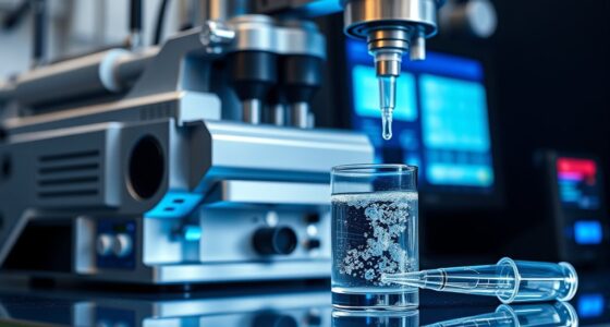
Have you ever wondered how scientists capture detailed images of tiny biological molecules? The answer lies in cryo-electron microscopy, or cryo-EM, a groundbreaking technique that allows us to see the unseeable. At the heart of cryo-EM is the process of preserving delicate biological samples in their natural state using rapid freezing. This step, called sample preparation, involves applying a small amount of the protein or complex onto a grid and then plunging it into liquid ethane at cryogenic temperatures. This quick freeze prevents the formation of damaging ice crystals, maintaining the sample’s structure with remarkable fidelity. Additionally, the sample preservation process is crucial for maintaining the biological activity and structural integrity of the specimen throughout imaging.
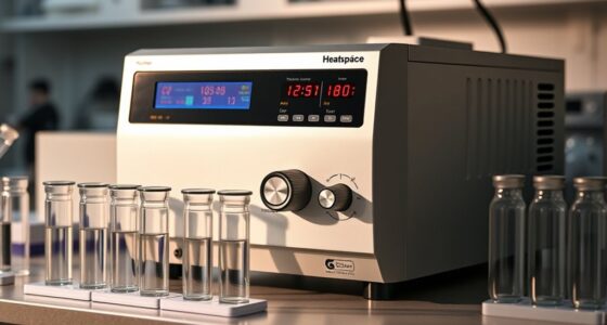
Once frozen, the samples are kept at cryogenic temperatures throughout imaging, guaranteeing they stay in their native conformation. The process of color accuracy preservation ensures that the structural details are captured with high fidelity, which is essential for accurate analysis.
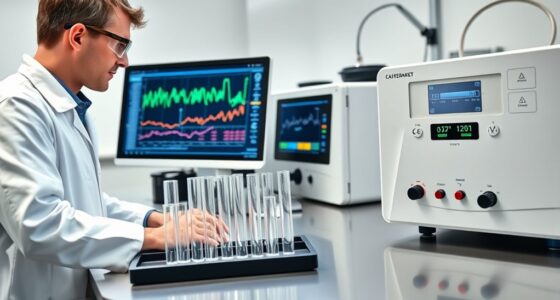
After preparing the sample, the next critical phase is data processing. You’ll use an electron microscope to shoot a series of two-dimensional images from different angles. These images are incredibly detailed but also noisy, so you need sophisticated computational tools to extract meaningful information. Data processing involves aligning thousands of these images, classifying similar views, and reconstructing a three-dimensional model of the molecule.
This computational step is vital because it transforms raw image data into a coherent, high-resolution structure. Advanced algorithms help to enhance the signal-to-noise ratio and refine the model, revealing the intricate features of proteins that were once invisible using traditional methods like X-ray crystallography.
Throughout this process, precision is key. Proper sample preparation ensures that particles are evenly distributed and oriented randomly on the grid, which is essential for accurate reconstruction. If the sample isn’t prepared correctly, the images may be blurry or incomplete, hampering the entire process.
Similarly, data processing depends on powerful software and algorithms that can handle massive datasets efficiently. These tools help you distinguish true structural details from noise, correct for sample drift, and improve the resolution of the final structure. The combination of meticulous sample preparation and advanced data processing enables cryo-EM to generate detailed, three-dimensional images at near-atomic resolution.
In essence, cryo-EM is a dance between careful sample handling and computational mastery. It’s about preserving biological molecules in their natural state through precise freezing and then leveraging digital tools to decode the complex data.
This synergy has revolutionized structural biology, allowing you to visualize proteins, viruses, and other biomolecules in unprecedented detail. By mastering sample preparation and data processing, scientists continue to reveal the secrets of life at the molecular level, pushing the boundaries of what we can see and understand in the microscopic world.
Frequently Asked Questions
How Does Cryo-Em Compare to X-Ray Crystallography?
You might find cryo-EM offers advantages over X-ray crystallography by requiring simpler sample preparation and avoiding the need for crystallization.
Cryo-EM provides high imaging resolution, allowing you to see proteins in their native states without extensive processing.
While X-ray crystallography can achieve higher resolutions with well-ordered crystals, cryo-EM is more versatile for complex or flexible samples, giving you detailed structures in more realistic conditions.
What Are the Limitations of Cryo-Em?
You might think cryo-EM has no limitations, but it does face sample limitations and resolution challenges. You need well-prepared, thin samples, and thick or complex specimens can be difficult to image.
While cryo-EM offers impressive insights, it sometimes struggles with achieving atomic-level resolution, making it less ideal for detailed structural analysis compared to other methods.
Recognizing these constraints helps you choose the right technique for your research.
Can Cryo-Em Analyze Live Cells?
You can’t use cryo-EM to analyze live cells because it requires rapid freezing, which halts cell activity. This limits your ability to study real-time cell membrane imaging and live cell dynamics.
Instead, cryo-EM provides detailed snapshots of frozen cells, capturing structures at a molecular level.
For observing live processes, techniques like fluorescence microscopy are better suited, as they allow you to track live cell behavior without freezing.
How Expensive Is Cryo-Em Equipment and Operation?
Imagine you’re setting up a cryo-EM lab, like researchers at a university. The equipment alone costs between $1 million and $3 million, with annual operation expenses reaching hundreds of thousands.
You’ll face significant cost analysis and funding challenges, as maintaining and running cryo-EM requires specialized staff and ongoing maintenance.
Despite these hurdles, the detailed insights gained make it invaluable for understanding complex biological structures.
What Future Advancements Are Expected in Cryo-Em Technology?
You’ll see that future cryo-EM advancements focus on overcoming automation challenges and improving sample preparation.
Expect more automated systems that speed up data collection and enhance accuracy, reducing manual effort.
Innovations may also streamline sample prep, making it more reliable and less time-consuming.
These improvements will make cryo-EM more accessible, allowing you to visualize even more complex biological structures with greater detail and efficiency.
Conclusion
With cryo-EM, you’re peering into the frozen silence of proteins, revealing their hidden dance in a delicate ice embrace. It’s like holding a whisper of life in your hands, turning the invisible into vivid detail. As you explore this icy window, you unlock secrets that once hid in shadows, revealing the intricate symphony of biology. Cryo-EM isn’t just a tool; it’s your key to seeing the unseen, alive and true.