
When inspecting particles with microscopes, understanding optical limits is essential for accurate results. Magnification alone doesn’t guarantee better detail; resolution, determined by factors like numerical aperture and light wavelength, sets what you can see clearly. Advanced illumination techniques improve contrast without surpassing these limits. Digital imaging enhances analysis but also relies on these fundamental boundaries. Knowing these optical constraints helps you avoid misinterpreting features and guarantees reliable insights—keep exploring to uncover more about optimizing your particle inspections.
Key Takeaways
- Optical limits, determined by numerical aperture and light wavelength, define the maximum achievable resolution in particle inspection.
- Magnification enlarges particles but does not necessarily improve detail resolution beyond optical constraints.
- Advanced illumination techniques enhance contrast and feature visibility without surpassing fundamental optical boundaries.
- Digital imaging extends effective resolution through high-resolution capture and pixel-based analysis.
- Proper tool combination and understanding of optical limits ensure accurate, reliable microscopic particle inspection results.
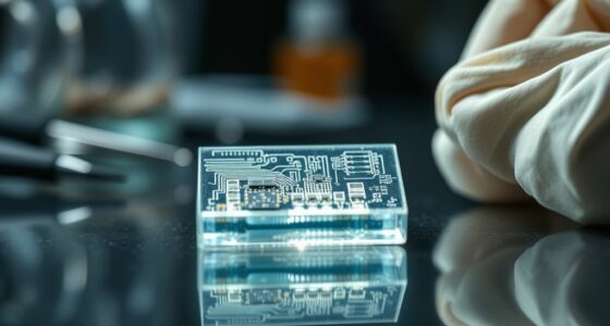
When inspecting tiny particles, having the right microscope can make all the difference. The quality of your results depends heavily on the tools you use, especially when it comes to resolving fine details. Optical limits are a critical factor in particle inspection because they define what you can see clearly and what remains beyond your reach. To push these boundaries, advanced illumination plays a vital role. Unlike traditional lighting, advanced illumination techniques, such as darkfield, phase contrast, or differential interference contrast, enhance contrast and reveal features that standard lighting might obscure. Proper lighting can help you distinguish particles against complex backgrounds, making subtle surface features or small contaminants more visible.
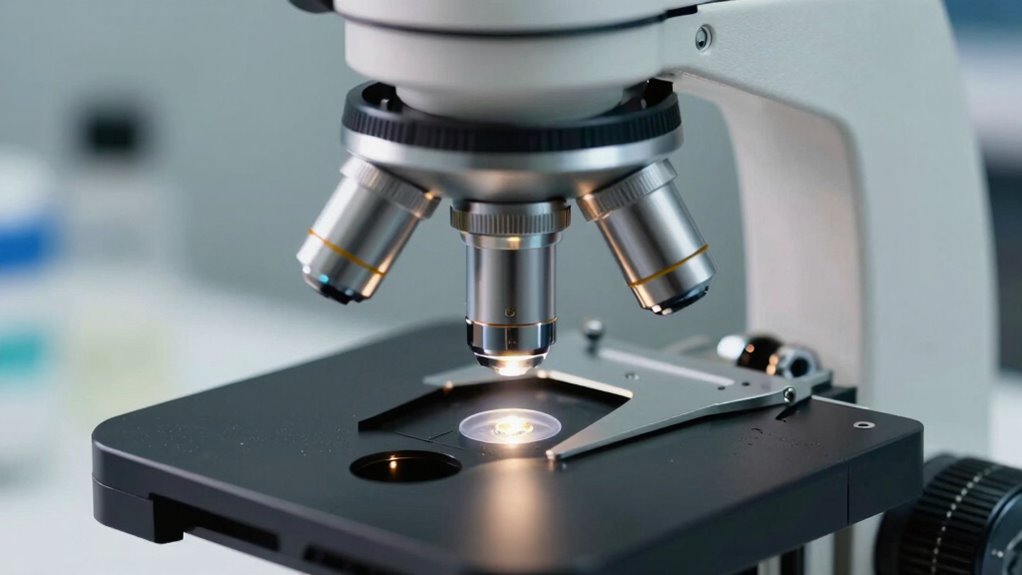
Digital imaging also significantly impacts your ability to analyze tiny particles. Modern microscopes equipped with high-resolution cameras let you capture detailed images, which you can analyze, share, or store for future reference. Digital systems provide the advantage of immediate feedback, allowing you to zoom in, measure, and compare particles precisely. This capability is especially useful when inspecting particles at the limits of optical resolution, as it helps you identify features that might be difficult to see with the naked eye or through traditional eyepieces. Additionally, digital imaging enables you to enhance images through software, improving clarity and contrast further without altering the actual sample. Understanding the optical limits of your microscope involves recognizing the role of magnification and resolution. While increasing magnification can make particles appear larger, it doesn’t necessarily improve your ability to distinguish closely spaced features. Resolution, determined by factors like numerical aperture and wavelength of light, sets the actual limit of detail you can observe. Advanced illumination techniques can sometimes improve effective resolution by enhancing contrast or reducing glare, but they don’t surpass the fundamental optical limits. That’s where digital imaging becomes invaluable—by allowing you to analyze images at a pixel level, you can extract more information than what’s visible through the eyepiece alone.
In particle inspection, you must balance various factors: choosing an appropriate magnification, employing advanced illumination to improve contrast, and leveraging digital imaging for detailed analysis. These elements work together to push the boundaries of what’s optically possible. Ultimately, understanding your microscope’s optical limits helps you make informed decisions, ensuring you get accurate, reliable results when inspecting tiny particles. With the right combination of illumination and digital technology, you can maximize your microscope’s performance and reveal details that might otherwise remain hidden. To optimize inspection outcomes, it’s also essential to understand optical limits, which define the boundaries of your microscope’s resolving power.
TOMLOV 4K Autofocus Microscope TM4K-AF Max, 10.1" HDMI Digital Microscope with Spin Flex Arm, Micro Soldering Microscopes for Electronics Repair, 52MP Error Coin Micro Scope Full View, Ring Light, 16G
Effortless Autofocus Microscope: The TOMLOV TM4K-AF Max delivers crystal-clear imaging with its advanced autofocus; Perfect for circuit repairs,…
As an affiliate, we earn on qualifying purchases.
As an affiliate, we earn on qualifying purchases.
Frequently Asked Questions
What Is the Maximum Particle Size Detectable With Optical Microscopes?
You can typically detect particles up to about 1 millimeter in size with optical microscopes. This is due to their particle resolution and magnification limits, which make larger particles easier to see clearly. As particle size increases beyond this range, detection becomes less precise because optical resolution drops. So, while microscopes excel at small particle inspection, they’re less effective for larger particles, which might require alternative imaging methods.
How Does Sample Preparation Affect Microscopic Particle Inspection?
Sample preparation greatly impacts microscopic particle inspection because it influences sample cleanliness and visibility. Proper techniques, like thorough cleaning and proper mounting, help eliminate debris that could obscure particles. If you neglect preparation, tiny particles might be missed or misinterpreted, leading to inaccurate results. By ensuring meticulous sample cleanliness and following effective preparation methods, you enhance the accuracy of your inspection and get a clearer, more reliable view of the particles present.
Can Optical Microscopes Differentiate Between Particle Materials?
Oh, of course, optical microscopes are just so advanced; they can effortlessly differentiate between particle materials, right? Well, not quite. You need spectral analysis to truly achieve material differentiation. Optical microscopes, limited by their optical limits, can sometimes distinguish color or shape, but identifying specific materials often requires more sophisticated techniques. Without spectral analysis, you’re mostly just guessing, making your inspection more art than science.
What Are Common Challenges Faced During Microscopic Particle Analysis?
During microscopic particle analysis, you often face challenges like sample contamination, which can obscure details and lead to inaccurate results. Additionally, limited image resolution hampers your ability to distinguish fine particle features, making material differentiation difficult. These issues require careful sample preparation and choosing the right microscope settings to improve clarity. Being aware of these common challenges helps you optimize your inspection process for more reliable and precise analysis.
How Do Environmental Conditions Impact Microscopic Inspection Accuracy?
Environmental factors like temperature fluctuations, humidity, and vibrations can markedly impact your microscopic inspection accuracy. These conditions may cause equipment drift or reduce image clarity, making calibration accuracy more challenging. To guarantee precise results, you should control the environment by maintaining stable temperature and humidity levels, minimizing vibrations, and regularly calibrating your microscope. Doing so helps you achieve consistent, reliable particle analysis and prevents errors caused by external influences.
darkfield microscopy for particle inspection
As an affiliate, we earn on qualifying purchases.
As an affiliate, we earn on qualifying purchases.
Conclusion
When choosing a microscope for particle inspection, understanding optical limits is essential. You’ll want to avoid the trap of thinking all microscopes can reveal every tiny detail—some particles are practically invisible to even the most advanced optics. By knowing these boundaries, you guarantee your inspections are both accurate and efficient. Remember, pushing beyond the optical limits is like trying to see the tiniest grain of dust in a hurricane—an exercise in futility that wastes your time and resources.
phase contrast microscope for tiny particles
As an affiliate, we earn on qualifying purchases.
As an affiliate, we earn on qualifying purchases.

10X Microscope Eyepieces Accessories for Optical Microscope Eyepieces Biological Microscopes Lens Adapters for Microscopes
High Quality: The optical microscope eyepiece is made of high-quality material and is durable and durable. Customers can…
As an affiliate, we earn on qualifying purchases.
As an affiliate, we earn on qualifying purchases.